
Our immune system relies on nanomachines, such as the membrane attack complex (MAC) to kill invasive bacteria in our blood. Our research, published in Nature Communications, provides us with a better understanding of how the immune system kills bacteria. This may guide the development of new therapies that harness the immune system against bacterial infections, and strategies that repurpose the immune system to act against other rogue cells in the body.

MAC pores imaged on the surface of a live E. coli bacterium - Heesterbeek et al, 2018
In earlier research, published in the EMBO journal, we showed that we were able to image the hallmarks of MAC attack, as pores on the surface of live bacteria, observing substructures such as the ‘stalk’ previously only visible by Cryo-EM.
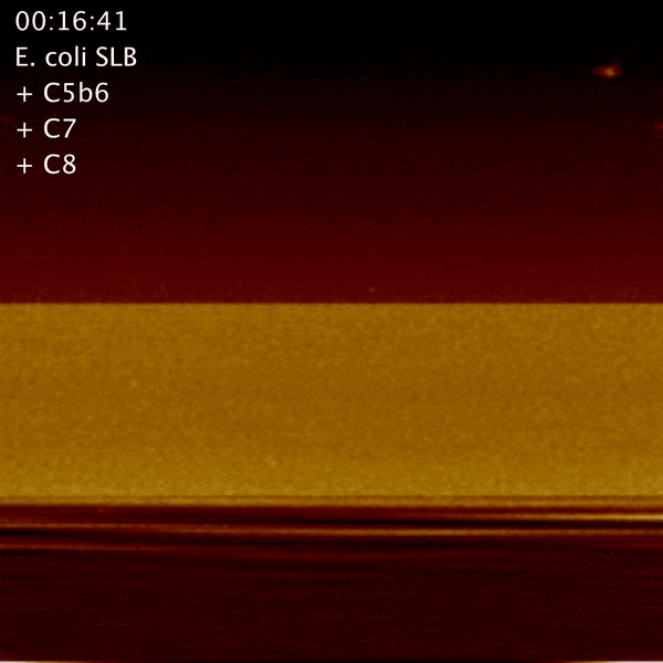
To determine the kinetics of MAC formation, we used high-speed atomic force microscopy (AFM), built and designed at the LBNI, EPFL, to track the process step by step.

The assembly of a MAC, imaged in real time - Parsons et al, 2019.
Interestingly, we found that there was a pause after the pore began to form, where the process stalled. The complex had partly formed, but was lacking over 50% of the complex, the part which forms the deadly pore. After the first of these missing proteins inserts into the bacterial surface, the rest fall into place much more rapidly, like dominos. Curiously, the main bottleneck – the insertion of this first protein – coincides with the point where hole formation is prevented on our own healthy cells, thus leaving them undamaged.
This research was led by University College London (UCL) and Imperial College London, in collaboration with the Swiss Federal Institute of Technology in Lausanne and the University of Leeds.