Experience of a Master's Student: Bradley Diggines

Every year we welcome undergraduate and masters’ students with a variety of backgrounds to undertake a research project in our lab.

What was your project about and what did it involve?

I was characterising the structure of kinetoplast DNA (a large network of interconnected DNA minicircles using atomic force microscopy and novel image analysis procedures. The project therefore had an even split of lab time, working on the Bruker Icon AFM, and programming, which built upon the TopoStats software.

What did you learn from your time in the lab?

I learnt a whole range of skills, both within the lab (I had never even used a pipette before!), to the computational programming side. As a materials science undergrad, these skills allowed me to transition into a more biological-chemical space. Alice also gave great feedback on my written/presented work which was very valuable.

What was the highlight/the best part about your time in the lab?

I particularly enjoyed using the AFM itself! While frustrating at times, when I finally saw new images appearing on the screen it was very satisfying. Generally, the lab was also a fun and supportive environment, with everyone willing to help out when needed :)

What are you doing now/any future plans?

I’m now a PhD student at Imperial College London, working on automated high-throughput microfluidic systems for the development of novel lipid nanoparticles and artificial cells. This work is in collaboration with AstraZeneca UK, aiming to transfect typically “difficult to transfect cells” within their pipeline. Supervised by Dr Yuval Elani, Prof Dame Molly Stevens and Dr Nick Brooks.

Good luck Bradley!

THE POWER OF AFM: AFM & SPM MEETING 2024

In March, the team visited the quaint city of Durham and attended the AFM & SPM Meeting 2024.

We started the exciting week off with a graduate school where we gained hands on experience on AFMs from various companies, a brilliant way to gain insight on the capabilities and advancements in AFM technology, in addition to lectures on the statistics for data analysis. The main conference revolved around AFM and SPM, covering a wide range of topics in Life and Biological Sciences, Materials Sciences and Energy, and Hardware, Software and Data Analysis. 

During the session on Life and Biological Sciences, Tom gave his talk titled “Determining DNA Damage by Atomic Force Microscopy”, providing insight on how powerful combining AFM with TopoStats, our program for automated tracing of biomolecules from AFM images, can be for advancing DNA topology research. His talk generated lots of interest from the audience!

Tom speaking at the Life and Biological Sciences session on “Determining DNA Damage by Atomic Force Microscopy”

Along with Tom’s talk, we had the privilege of attending numerous talks that captivated our attention with their thought-provoking content. We found ourselves deeply engaged and inspired by the diverse range of topics that were covered, leaving each of us with new ideas and perspectives to ponder. Amongst the speakers was Wei Feng Xue who performed structural analysis of helical filaments using three-dimensional contact point reconstruction AFM and Paolo Actis who demonstrated how AFM can be used for single molecule delivery into living cells.

We finished the days off with a poster session that bridged researchers from various fields, enabling us all to engage in in-depth discussions and broaden our knowledge on the potential of AFM in a plethora of research fields. During these sessions, Libby, Sylvia, Tobi and Laura were able to present their research to the community, allowing them to exchange insight with other researchers and provoke fruitful discussions on their respective research projects. 

It came as no surprise during the conference dinner when Sylvia and Libby were awarded the 1st and 3rd prize, respectively, for their outstanding poster presentations, a well-deserved recognition from their peers and the judging panel.

Sylvia (left) and Libby (right) awarded with the 1st and 3rd poster prizes, respectively.

The conference was both enjoyable and enlightening. Engaging with cutting-edge research, networking with fellow researchers and participating in thought-provoking conversations have enriched our understanding and inspired our research moving forward.

Max tackles the topographically complex mountains of banff with topology 2024

In March, Max visited the Rocky Mountains of Banff national park to attend the 2024: The Crossroads of Topology, Combinatorics and Biosciences: Deciphering the Entanglement of Multi-Stranded Nucleic Acids workshop

Max giving his presentation on new image analysis methods to characterise DNA Topology

Set within the heart of the Banff national park, this 5-day conference brought together researchers from a range of fields to remove our blinders and open our eyes to the multitude of research attacking the same topological problems from various angles. From hardcore mathematical modellers developing language models to probe the intricacies RNA structures, to the dedicated biologists investigating ion interactions on  and computational modellers, this workshop was a great opportunity to merge these fields and push the understanding of DNA topology.

At the workshop, I heard some truly amazing talks surrounding the multitude of DNA structures in various environments, and how this structure can regulate access to the genome, as well as insight into the models and physical laws used to govern them. Standout talks include Lynn Zechiedrich showing negative supercoiling induced DNA-protein interactions occur at a supercoiling threshold, alongside fruitful results surrounding supercoiling induced defects from a past collaboration with Alice. I was also intrigued by Andrew Rechnitzer research into streamlining pivot algorithms to quickly model complex knot and catenated lattice. I also wanted to shout out the superb talk given by Lila Kari where she used machine learning to identify clusters of taxonomic and environmental factors from fragmented DNA sequences of various species.

Group photo of conference attendees

There were many insightful discussions to highlight the biological questions faced by the community such as how does DNA packing affect genome access? It gave a chance for people to spotlight their research which pushed towards this goal, and discussed the ways in which the expertise from these different fields could be utilised to move together towards the biological answers we seek via developing collaborations.

*Most talks should be available on the BIRS website here

UK SPM/AFM Users Meeting 2024

In January 2024, the lab attended the Bruker UK SPM/AFM Users Meeting 2024. It was a privilege to attend the event which was held at the Henry Royce Institute in Manchester. The days were split into two with demonstrations of incredible microscope technology in the morning followed by an afternoon of fascinating, frontier pushing science. As single molecule, bio-AFM users it was particularly interesting to hear about developments in other SPM technologies. For example, AFM-IR which allows the operator to gather information about their sample surface as well as infrared spectra helping identify the materials it contains and it was intriguing to think about how this could be applied to our work.

Two standout talks came from our very own Max Gamill, who described recent advances in TopoStats’ ability to resolve topology from AFM images and Dr Laia Pasquina Lemonche, who discussed her work developing software to study bacteria with the aim of developing new antibiotics, which was thought provoking and very exciting.

In the evening, we attended a meal where it was an absolute pleasure to listen to Dr Alex Ramadan tell the story of her journey to academia. The food was amazing and one of the highlights of the evening was the AFM quiz, which caused an unexpected explosion of competitiveness between tables. 

Alice presenting our work on automating tracking and tracing of DNA

Overall, the conference was a blend of mind -blowing science and discussions with enthusiastic microscopists! We’re looking forward to next years event.

Taking AFM to the Alps- EMBO DNA Topology Workshop

In September, Max, Libby, Eddie, Tom, and Alice visited the picturesque Swiss village of Villars-sur-Ollon to attend the 2023 EMBO DNA topology and topoisomerases in genome dynamics workshop. 

Set against the beautiful backdrop of the Alps, we first attended an LS2 ECR satellite event (chaired by Alice) where Eddie, Libby, Max and Tom all gave a 5 minute flash talk introducing their research. This event also gave us all an opportunity to present our research and network with our fellow ECRs while getting to know the scenic setting for the conference. 

Max speaking at the ECR satellite events about his research using new image analysis methods to characterise DNA topology

The main conference featured fantastic talks covering the critical role of DNA topology in a wide range of processes, offering deep insights into its implications for transcription regulation, topoisomerase activity, as well as its impact on various aspects of disease progression and developmental biology. Standout talks included a captivating keynote by Tony Maxwell, delving into his personal journey in topoisomerase research. His insightful anecdotes and scientific contributions illuminated the evolution of this field. Additionally, Valerie Lamour delivered a compelling presentation on the binding mechanisms of DNA gyrase to supercoiled DNA and its implications in DNA biology.

During the Regulation of DNA Topology and Topoisomerase Activity session, Alice delivered her talk titled "Tackling Topology with TopoStats." It was well reived by the audience and provoked interesting questions and lead to lots of discussion in the coffee sessions and at dinners that followed.
The poster sessions saw Max, Libby, Eddie, and Tom showcase their work which inspired lots of thought-provoking questions about the potential of AFM and advanced image analysis techniques in DNA topology research and led to productive discussions about future work and collaboration.

Alice speaking at the main workshop on “Tackling Topology with TopoStats”

The trip allowed the those of us attending from the Pyne group to reflect on our research within the broader context of the field and plan the upcoming stages of our research. The conference provided an invaluable platform for networking and engaging in in-depth discussions on our research in DNA topology, bringing together researchers at different career stages from across the field. The anticipation is that these discussions will pave the way for fruitful research and collaborations in the future.

Eddie (top left), Max (top right), Tom (bottom left) and Libby (bottom right) presenting their posters at the main conference poster session.

New PDRA position in experimental DNA biophysics

We are starting off 2024 with the exciting news that we have an open PDRA position in our group, fully funded by Alice’s UKRI Future Leaders Fellowship. This position would suit someone with an interest in single molecule microscopy, (bio)image analysis, DNA biophysics or therapeutic development.

Everyone is looking for great postdocs at the moment, so here's a little on who we are, who we're looking for and what I think we can offer:

  • We are a really close group, united by interests in open science, microscopy, DNA structure and bioimage analysis.

  • We work together as a group and with amazing collaborators to try and solve interesting problems at the interface of the physical and life sciences.

  • We believe in balancing our work and personal lives, and taking time to enjoy all that Sheffield and the Peak District has to offer.

  • We're looking for someone who is excited by our research and working with us to understand the role of DNA’s mechanical code in its interactions.

  • We don’t need you to have very specific skills which we can teach, but to have an open mind, an interest in DNA biophysics, open science and single molecule microscopy.

  • We're really happy to support you in applying for grants/fellowships/permanent roles - we have experience in all of these and will support you in doing so.

This position is for around 2.5 years (until 31 July 2026) with the potential for renewal if you're enjoying the role.

The job description is here, and please get in touch if you think you'd be interested in working as part of our team. If you don't have all the criteria, or aren't sure if this is the right job for you, let's have a chat and see!

Experience of a Masters Student: Chloe Moor’e

Every year we welcome undergraduate and masters’ students with a variety of backgrounds to undertake a research project in our lab. Chloe Moore completed her MRes in Translational Oncology in 2022 where she was looking at the damage caused by different forms of radiation on DNA by using AFM in the Pyne lab.

What was your project about and what did it involve?

My project involved comparing the different types of DNA damage/lesions induced by both gamma and alpha radiation. I did this by exposing both MDA-MB-231 cells and piAN7 plasmids to different doses of gamma radiation and alpha radiation (radium dichloride). Gamma and alpha radiation cause DNA damage via different mechanisms therefore finding an equivalent dose is impossible. However, using cells allowed me to carry out clonogenic assays to determine a rough estimate for an equivalent dose of gamma and alpha radiation by seeing which doses of each type induced the same amount of death. As piAN7 plasmids are circular in nature, they exhibit the different types of DNA lesions impeccably and therefore allowed me to characterise the lesions and determine which type of radiation induced which types of damages.

What did you learn from your time in the lab?

I learned that things don’t always go to plan, and you must be persistent and resilient to succeed and make progress. You must be adaptable to overcome any mishaps that may occur.

What was the highlight/the best part about your time in the lab?

The highlight of my time is the lab was definitely the people I met. My lab group were always so helpful, and they always made sure they made time for me if I had any questions or needed any help. Although completing my masters was very academically challenging, everyone is always up for a social event or even just taking a break and grabbing a coffee to help wind down.

What are you doing now/any future plans?

Currently I am working as a healthcare assistant at a local GP practice. I am however, browsing for jobs linked to medical sales and clinical research so that I can kickstart my scientific career.

Physics of Life 2023

In March, Libby, Eddie, Max, Tom, Laura, and Alice all attended the Physics of Life conference at Harrogate Convention Centre. This was the first conference of its kind, representing science at the interface between biology and physics.

During the conference there were keynote lecturers and parallel sessions covering a wide range of topics from single molecule biology to evolution, as well as two poster session and a conference dinner allowing plenty of opportunities for networking.

There were many excellent talks, but the standout talks included hearing Graeme King from UCL discuss the mechanism of supercoil relaxation by the Human Topoisomerase3α RMI1-RM2 complex and Sarah Harris from the University of Leeds discuss how her simulations can give insight into how molecular motors “walk”.

Alice speaking at the Physics of Life roadmap session

Alice presented during the Single Molecule biology session on “Tackling Topology using TopoStats” and also was part of the Physics of Life roadmap panel where she advocated for better support of early career researchers. Laura presented during the Machine Learning at the Physics/Biology interface session on “CellPhe: a toolkit for cell phenotyping using time-lapse imaging and pattern recognition” discussing the work she has conducted during her PhD at the University of York. Both talks were greatly received with lots of interest from the audience. 

Laura presenting on her PhD work- CellPhe

Additionally, Libby, Max and Eddie presented posters about their work. These posters provoked lots of interesting discussion and questions during the poster session.

Eddie, Max and Libby presenting their posters.

The conference was an excellent opportunity to engage with research from across biology and physics as well as a chance to share and discuss the research we have been doing in our lab with the wider biophysics community.

UK Bruker Users Meeting- Leeds 2023

In January Tom, Rob and Eddie went to the 2023 Bruker UK AFM user meeting in the Bragg Centre at the University of Leeds. This was the first user meeting that Bruker has run in three years due to the COVID-19 pandemic. Over two days we were able to attend practical workshops and see demonstrations of the latest developments from Bruker’s AFM division. We also heard a great selection of talks from AFM users from across the UK working in a range of different disciplines.

Highlights included hearing Ioanna Mela from Cambridge talk about how AFM can be corelated with super-resolution microscopy to study bacterial cell membrane components in supported lipid bilayers and then seeing a demonstration of the new high-speed NanoRacer system in action.

Eddie also had the opportunity to presented a poster on his work on the interaction of NDP52 with DNA and won the second place poster prize.

Eddie and his poster

Eddie receiving his poster prize

The meeting was a good opportunity to meet and discuss AFM with researchers from around the country, both in the scheduled sessions and the conference dinner where we also put our general and AFM knowledge head to head in a fun quiz.

Research Software Engineering Conference 2022

From the 5th to the 8th of September 2022, Sylvia (writing) with Neil and Bob from the Sheffield RSE team, attended RSECon2022 a conference centred around Research Software Engineering (RSE) in Frederick Douglass Centre at Newcastle University.

RSECon2022 brought together, in Newcastle, people from around the world interested in developing software to support and facilitate scientific research. Talks and panels were wide ranging, from computational and programming methods to discussions on how the RSE community can be more inclusive. I found all the talks that I attended to be both very interesting and informative, especially the ones discussing the different experiences and kinds of work that research software engineers do.

An important thread that ran throughout the conference was ensuring good and open research through the FAIR principles (Findable, Accessible, Interoperable, Reproducible) which has been an important consideration in my own work and it. These themes were discussed at length, with a lot of enthusiastic community engagement in the form of questions and proposals from the audience.

The technical talks were also wide ranging and helpful, I learned a lot about new and cutting-edge software while also gaining more understanding of the software development process for small to medium teams in RSE.

“Make your code 10,000 times faster with parallel numpy” was an introduction on using tools like vectorisation and numba to significantly decrease computational times in workloads, “Quarto - a library to run them all?” showcased Quarto, a platform agnostic, multi-language system for scientific and technical publishing that can be used for making documents such as interactive and dynamic presentations with inbuilt code blocks (and running code!), rendering, and even maps. “Global.health - lessons from building a data platform during the pandemic” was a dive into the difficulties of building critical software infrastructure for governments and health agencies during the pandemic, and the fast-paced nature of writing software during rapidly changing circumstances and needs.

I’m planning to do a software and computationally focused PhD in the near future so this conference allowed me to widen my understanding of research software engineering, find out what kinds of things people do in the field and network to get to know people who may have experience of what I want to do in the future. These discussions with people from different RSE teams and backgrounds was helpful in understanding my options going forward, as well as what it is like to do a computationally focused environment and the opportunities that may come after a PhD.

I cannot overstate how useful and enjoyable the experience was and I look forward to attending more conferences and events like this in the future

Experience of a Masters Student

Following on from our pervious blog post about how Eddie transitioned from working at AFM probe manufacturer NuNano to doing a PhD in the Pyne lab, this blog post looks at the experience of Billy Davies in the lab before he made the reverse journey from Sheffield to Bristol.

Billy completed a project masters project in the Pyne lab in 2021 as part of his MSc in Molecular Medicine at the University of Sheffield and now works at NuNano in Bristol as a probe consultant.

Billy Davies is now a Probe Consultant for NuNano

What was your project about and what did it involve?

My project studied how DNA topology affected on the binding of DNA Gyrase and its subunits. Gyrase is an essential enzyme involved in relaxing over-twisted DNA, and detangling DNA loops. We wanted to see if different conformations and sites where DNA strands crossed over affected the binding preference of Gyrase. Using AFM, we imaged a variety of DNA substrates, some overwound DNA and others linked rings, with and without Gyrase and its subunits, so we could see if there were preferences for some conformations over others.

What did you learn from your time in the lab?

My time in the Pyne Lab can be summed up by the fact that I came in having heard the technique in passing and left an AFM believer, taking my first job at NuNano (an AFM probe manufacturer). Everyone in the group is so enthusiastic about AFM it was hard not to end up finding it fascinating, even if its completely different to what I had studied in the past, and I really felt like I grew as a scientist by working with them. Despite the restrictions, the amount of effort they made to get me trained and on the microscope was amazing and I’m incredibly grateful to have had the opportunity to learn from leading AFMers such as Alice.

Summarise your experience in the lab

The best thing about being there was how much effort they made to make you feel like a member of the group, and that your project (even if it was one of many) was important for the lab as a whole. Alice was always interested in what we were doing, and listened to any of my ideas or plans even if they weren’t actually very good, and gave us just the right amount of support vs independence.

Billy at the AFM

Billy on the AFM at NuNano

Selling probes to pursuing a PhD- Eddie's move, one year on

This blog post originally appeared on the NuNano website where Eddie sat down (virtually) with his former colleagues to discuss his move from selling AFM probes in Bristol to starting a PhD using AFM in Sheffield. You can see the original post here.

After nearly 2 years working with NuNano as our Sales Coordinator, engaging with lots of scientists using AFM, Eddie Rollins found the lure of academic research too strong to resist.

Eddie made his return to university last November to undertake a PhD with Dr Alice Pyne at the University of Sheffield and we caught up with him earlier this month to find out how he was getting on, one year into his studies.

Eddie Rollins at work in the nanocharacterisation lab in the Royce Discovery Centre.

Great to catch up with you again Eddie! Can you tell us a bit about your PhD?

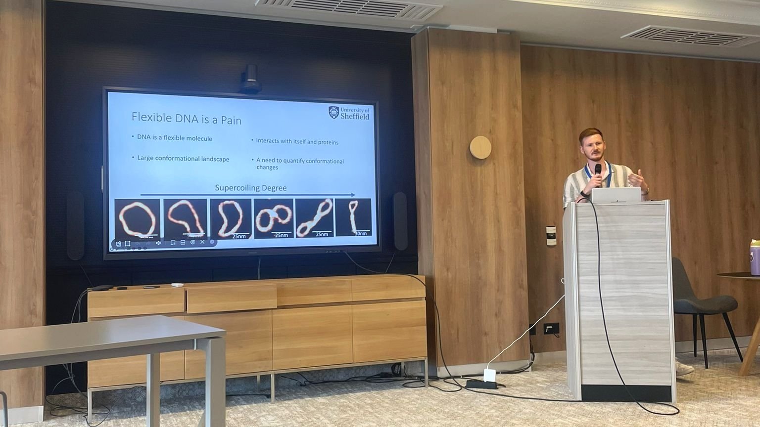
Sure. I’m trying to understand how small-scale changes in the structure of DNA affects how it interacts with proteins.

These interactions are biologically essential for all forms of life so are important targets for antibiotic and anti-cancer drugs. Understanding how they work could help develop and improve treatments for a wide range of diseases.

AFM is an essential tool to help understand these interactions since it allows us to see individual molecules of DNA and structural changes such as bends and twists as well as directly visualizing the DNA and protein binding.  

In what way are you using AFM? To what extent did your background of working with NuNano help at all?

AFM is a large part of my PhD. I use AFM to image individual molecules of DNA as well as their interactions with proteins. I take advantage of AFMs ability to operate in liquid to get high resolution images of these interactions in a near native state which allows me to see how the local shape of the DNA is affected.

Working with NuNano was a huge help when I started doing AFM as I’d already learnt a lot about the technique from the NuNano team, as well as through discussions with customers about their work in a wide variety of fields.

Additionally, as the probe is such an core part of the microscope, having a good grasp of the different aspects of probe design made a big difference not only in understanding probe choice in my own lab but also in evaluating AFM experiments in the literature.

What was it like moving from working with NuNano to working back in academia again?

Doing a PhD is a long game, the deadline is 4 years away at the start, so you have to create your own structure and your own intermediate targets.

While at NuNano I had a constant stream of calls and meetings with customers on top of the regular meetings and deadlines that come with being part of a growing company. This was probably the biggest adjustment.

Whilst I’ve enjoyed the freedom and space to do research and learn I do sometimes miss the faster-paced world of commerce and chasing sales!

How was it moving to a new area and starting a new job whilst we were still very much in the full throes of the Covid pandemic last autumn?

Moving during a pandemic was pretty difficult. Not only the practical side of it was hard but it’s taken a while to get to know the city and people in it. Thankfully Sheffield is a very green city with easy access to the Peak District so I manged to discover a lot of lovely parks and explore the Peaks on my bike.

The PhD itself also didn’t start as I had imagined as I wasn’t able to get into the lab for four months - though that did give me a lot of time to read and attend the glut of virtual conferences going on at the time.

Although it was a bit frustrating at the time it meant that when I did get into the lab I had a lot better idea of what I was doing and was able to hit the ground running and get some good data, so it worked out well really.

As an insider to academia now, what are your thoughts on the need and value of an AFM Community?

AFM is a powerful technique with potential applications in numerous areas from nanotechnology to diagnostics. This breadth of application has the potential to fragment the field so having a strong and generous AFM community is essential.

Sharing developments and best practice between people and labs working in different areas through such a community is one way to ensure that all forms and applications of the technique can benefit from these improvements.

I believe an AFM community that involves the commercial instrument and probe manufacturers is key to seeing the applications developed in academic labs applied more broadly in non-specialist setting like the clinic.

What’s brilliant is that since starting my PhD I’ve seen the AFM community come together around the question on data analysis and seen projects emerge to share software between groups (see www.github.com/AFM-SPM). I can see this being a particular area of collaboration allowing progress like we’ve seen in the cryo-EM field.

What has been the biggest lesson for you in the past year?

I think one of the biggest lessons I’ve learnt this year is that doing AFM provides fantastic opportunities to collaborate with a variety of researchers.

Being able to directly view nano-scale structures can clarify mechanisms that otherwise can only be visualised abstractly as cartoons so combining AFM with other approaches can reveal things invisible to each technique in isolation.

This is true of all scientific instruments and methods however AFM’s fairly unique set of advantages and disadvantages makes it a particularly helpful partner for illuminating processes at the nanoscale. 

Eddie and James Vicary (NuNano MD) at low-key COVID send-off for Eddie after 18 months working for NuNano.

Paper published showing the helix of ‘dancing DNA’

Alice and Kavit alongside collaborators have published a paper in Nature Commmunications this week, ‘Base-pair resolution analysis of the effect of supercoiling on DNA flexibility and major groove recognition by triplex-forming oligonucleotides’.

Videos accompanying the paper, derived from high resolution AFM images of DNA and molecular dynamics (MD) simulations, show for the first time how small circles of DNA adopt dance-like movements.

 
DNA minicircle wiggling.
 

The footage is based on the highest resolution images of a single molecule of DNA ever captured. They show in unprecedented detail how the stresses and strains that are placed on DNA when it is crammed inside cells can change its shape.

Previously scientists were only able to see DNA by using microscopes that are limited to taking static images. But now the Yorkshire team has combined advanced atomic force microscopy with supercomputer simulations to create videos of twisted molecules of DNA.

Alice said:

 “Seeing is believing, but with something as small as DNA, seeing the helical structure of the entire DNA molecule was extremely challenging. 

The videos we have developed enable us to observe DNA twisting in a level of detail that has never been seen before.”

The images are so detailed it is possible to see the iconic double helical structure of DNA, but when combined with the simulations, the researchers were able to see the position of every single atom in the DNA and how it twists and writhes. 

Every human cell contains two metres of DNA. In order for this DNA to fit inside our cells, it has evolved to twist, turn and coil. That means that loopy DNA is everywhere in the genome, forming twisted structures which show more dynamic behaviour than their relaxed counterparts.

The team looked at DNA minicircles, which are special because the molecule is joined at both ends to form a loop. This loop enabled the researchers to give the DNA minicircles an extra added twist, making the DNA dance more vigorously. 

When the researchers imaged relaxed DNA, without any twists, they saw that it did very little. However, when they gave the DNA an added twist, it suddenly became far more dynamic and could be seen to adopt some very exotic shapes. These exotic dance-moves were found to be the key to finding binding partners for the DNA, as when they adopt a wider range of shapes, then a greater variety of other molecules find it attractive.  

Previous research from Stanford, which detected DNA minicircles in cells, suggests they are potential indicators of health and ageing and may act as early markers for disease.

As the DNA minicircles can twist and bend, they can also become very compact. Being able to study DNA in such detail could accelerate the development of new gene therapies by utilising how twisted and compacted DNA circles can squeeze their way into cells. 

One of our collaborators who produces the minicircles used in this study said:

“Dr. Pyne and her co-worker’s new AFM structures of our supercoiled minicircles are extremely exciting because they show, with remarkable detail, how wrinkled, bubbled, kinked, denatured, and strangely shaped they are which we hope to be able to control someday.”

You can read the paper here:

Base-pair resolution analysis of the effect of supercoiling on DNA flexibility and major groove recognition by triplex-forming oligonucleotides

Isabel and Alice publish paper on a new way to detect antimicrobial resistance

A new, quicker way of detecting antibiotic resistance in bacteria has been developed by a team of scientists from the EPSRC funded interdisciplinary research collaboration, i-sense.

Isabel Bennett and Alice Pyne along with collaborators at UCL have developed a new technique that uses nanotechnology to detect antibiotic resistance in approximately 45 minutes.

bennett.gif

The standard method for detecting resistance is a relatively slow process that typically takes between 12 and 24 hours. The ability to reduce this time could significantly help the ongoing battle against antibiotic-resistant bacteria - a problem which is predicted to cause 10 million deaths per year and cost the global economy $100 trillion by 2050.

Speeding up the time it takes to identify antibiotic-resistant bacteria could improve our ability to prescribe antibiotics correctly and reduce the misuse of antibiotic treatments - a key step in the fight against antibiotic resistance.

The new method was developed by Isabel at UCL in collaboration with Alice and Prof. Rachel McKendry, also from UCL, and uses a new Atomic Force Microscopy (AFM) detection system. 

Our method allowed us to quickly differentiate between resistant and sensitive phenotypes in multiple strains of E. coli, a bacteria implicated in a number of challenging infections including UTIs.

Dr Isabel Bennett

UCL

This method uses a nanomechanical cantilever sensor together with a laser to detect single bacterial cells as they pass through the laser’s focus, which provides a simple readout of antibiotic resistance by detecting growth (resistant) or death (sensitive) of the bacteria.

By placing a reflective surface - a small stiff cantilever - in a filtered growth medium in a petri dish and reflecting a laser off it onto a photodiode detector, it is possible to detect bacteria as they pass through the path of the laser, therefore altering the signal at the detector.

Following the addition of the antibiotic to the petri dish, the study has shown that it is possible to detect whether fewer bacteria interfere with the laser beam, thereby indicating cell death in the antibiotic-sensitive bacteria.

The new technique developed by Isabel builds on an AFM method from a previous study, however this method doesn’t require the bacteria to be immobilised - making the new detection system much faster.

Isabel said: “Our method allowed us to quickly differentiate between resistant and sensitive phenotypes in multiple strains of E. coli, a bacteria implicated in a number of challenging infections including UTIs.”

Alice added: “We were able to show that our faster method was able to reproduce values from gold standard measurements, such as MIC’s in a fraction of the time.”

The study - Cantilever Sensors for Rapid Optical Antimicrobial Sensitivity Testing - was conducted by Dr Isabel Bennett as part of her PhD supervised by Dr Alice Pyne and Professor Rachel McKendry.

The research by the all-female team of scientists is published in the journal ACS Sensors. The journal has published an interview with Dr Bennett following the paper being selected as an ACS editors choice.


Read the paper here

EMBO DNA topology and topoisomerase meeting in Les Diablerets

EMBO DNA topology and topoisomerase meeting in Les Diablerets

I was lucky enough to have been selected to give a talk at the 2019 EMBO workshop on DNA topology and topoisomerases in genome dynamics in Les Diablerets. This was my second time attending this meeting, and it was particularly exciting for me, as I was returning to present data that came from a new collaboration with James Provan, Sean Colloms and Andrzej Stasiak which came from the last EMBO meeting.

Isabel passes her viva! Congratulations Dr Bennett

Isabel passes her viva! Congratulations Dr Bennett

Congratulations to Dr Bennett on passing (and enjoying) her viva, and thank you to her brilliant examiners, Til Bachmann and Carmel Curtis for making it such a great experience. Its been an absolute pleasure working with Isabel for the past 5 years - I’m excited to see what the future has in store for you.